The brain carries endless mysteries and unlimited potential. It is the most complex and mysterious part of the human body and has always been the focus of scientists’ exploration. This delicate organ not only controls our behavior, but also deeply affects our emotions, thinking and memory. However, our understanding of the brain has so far been only the tip of the iceberg.
From May 13 to 14 (Beijing time), Tianqiao and Chrissy Chen Institute (TCCI) and NeuroPSI organized an international conference themed “Brain, Behavior & Beyond”.
On the first day of the conference, five renowned scientists in the field of brain sciences gave a lecture on brain development and sensory cognition. On the second day, four renowned scholars, including Nobel Laureate Professor Michael Rosbash, enthusiastically shared the latest research progress in the areas of circadian rhythms, emotions, and strokes.
TCCI has live streamed the conference online to allow more scholars to participate in the scientific event first-hand.
Tagging circadian neurons to edit sleep?
With summer approaching, we occasionally see a few annoying fruit flies on the fruit we forgot to eat in time. However, this rice-sized creature is a favorite in scientific research. As a model organism, the fruit fly is widely used in the study of biological rhythms.
Professor Michael Rosbash from Brandeis University in the United States has used fruit flies to study circadian rhythms, a pioneering work that earned him the Nobel Prize in 2017. In this session, Professor Rosbash talked about how to incorporate new technologies for studying the circadian rhythm of fruit flies.
As a new method for assessing the early POI-II activities in RNA transcription, BuTT-Seq can record transcriptional regulation of RNAs. It is faster and more efficient than the established Nascet-Seq and NET-Seq, which provides a time window for observing the mRNA of the core clock gene of fruit flies, which has a short maintenance time of activity. In addition, the eRNA identified by BuTT-Seq is an enhancer that has not yet been defined and is hypothesized to exist only in clock neurons.
The brain of a fruit fly possesses between 10,000 to 30,000 cell types, and the multitude of cell types provides the cellular biological basis for behaviors such as circadian rhythm, sleep, learning memory, fighting, motivation, mating and courtship, and addiction. The circadian behaviors of fruit flies are regulated by a variety of neuronal subtypes. Regulator genes, and genes related to neural connectivity molecules are the most abundant molecular markers for cell subtype clustering. Meanwhile, single-cell sequencing (scRNA-seq) can further type the circadian neurons and thus discover the correlation of marker gene expression.
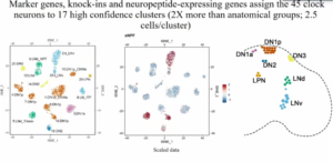
During the meeting, Professor Rosbash pointed out that there are very significant cell surface molecules in the biological clock system of adult individuals, which specifically interact with each other to regulate the specific firing of neurons during development. The expression of cell surface molecules is neuron-specific. For example, G protein-coupled receptors can be a good marker for neuronal typing, which is important for the maintenance of neural circuits and neuroplasticity.
In the end his presentation, Professor Rosbash concluded that scientific progress relies on new technologies, methods, and ideas, and the emergence of new technologies paves the way for new methods and ideas.
Mitochondrion is the culprit responsible for depression and anxiety?
Living in a fast-paced world, have you experienced the same feelings of anxiety due to approaching deadlines? Have you chosen to give up instead of working hard as a result of heavy social stress? These experiences encouraged researchers to embark on research into how human brains encode negative emotions. Professor Carmen Sandi from the Swiss Federal Institute of Technology in Lausanne talked about the mechanisms behind the engagement of mitochondria in regulating anxiety and motivation from the perspective of brain circuits.

Professor Sandi selected rats as the experimental subjects and made them anxious through social competition experiments and used molecular biology techniques and electrophysiological recordings to reveal the differences in the activity of NAc neurons in the low-anxiety group and high-anxiety group. She further used rescue, overexpression, and inhibition experiments to demonstrate that the mitofusin-2 (MFN-2) in the MSN-D1 and MSN-D2 neurons in the NAc brain region is necessary for the structural and functional alterations of the MSN neurons in the different anxiety groups.
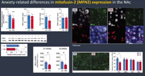
Finally, Professor Sandi shared unpublished research findings. The research team used transgenic technology to compare the functional differences of MFN-2 in MSN-D1 and MSN-D2 neurons, to further validate the effect of limbic system modulation of anxiety on motivation, and the alteration of blood-brain barrier by stress and anxiety.
Based on these findings, Professor Sandi proposed the neural encoding hypothesis of mitochondria and metabolism of stress-coping/motivational behaviors, that is: altered mitochondrial MFN-2 levels — abnormal structural function of specific neurons — altered behaviors, which has been validated by human experiments and experiments on the hippocampus of rats.
Sleep consumes energy as well; energy metabolism is modulating sleep?
The daytime and night are two parallels whose paths will never cross. Some people are full of energy during the day, while some tossing and turning late at night. Sleep is an instinctive behavior of human beings that evolved from living with nature and is regulated by biological rhythms. Professor Gero Miesenböck from the University of Oxford shared the microscopic mystery of sleep disorders from a biological perspective.
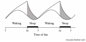
Sleep is regulated by specific neurons whose electrical signals are altered when there is pressure to fall asleep, and waking is influenced by the activation of the dopamine system. So, the researchers wondered, what causes the stress of falling asleep? And what regulates waking and sleep?
It was found that sleep stress is associated with the loss of carbonyl reductase. Carbonyl hydroxyl compounds originated from polyunsaturated fatty acid are signaling intermediates that couple mitochondrial electron transport to sleep and convert Kvβ to NADP+ channels. The loss of carbonyl reductase prevents these processes from operating properly.
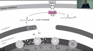
Professor Miesenböck further elaborated on the microscopic processes of energy metabolism during waking and sleep and how it interfaces with ion channels and affects sleep. Kvβ enhances sleep by sensing redox reactions during cellular energy metabolism, and this regulation is achieved by controlling the activity of sleep-associated neurons. In contrast, sleep deprivation impairs the lipid oxidation chain reaction, which in turn affects the sleep-enhancing attribute of Kvβ.
In the end, Professor Miesenböck concluded that sleep is not only related to metabolism, but also involved in memory consolidation and even exerts influence over the lifespan of an individual.
Small vessels harbor big problems; Is stroke preventable?
In the microscopic world, a mutation of a gene and the abnormal function of certain cells may cause severe health issues such as vascular obstruction or rupture. During the meeting, Professor Anne Joutel from the Institut de Psychiatrie et Neurosciences de Paris, shared the latest research progress on the occurrence of cerebral small vessel diseases (CSVD).
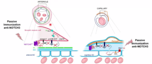
Professor Joutel began by describing how abnormal cerebrovascular events cause blood flow variability in the brain when CSVD occurs. To facilitate the presentation, she chose to share a highly prevalent CSVD: CADASIL (cerebral autosomal dominant arteriopathy with subcortical infarcts and leukoencephalopathy). A distinctive feature of CADASIL is a site mutation of the NOTCH3 gene when encoding cysteine. A mouse model suggests that early onset of CADASIL is characterized by an increase in the cerebral small vessel basilar membrane NOTCH3ECD and the deposition of granular oleophilic material GOM, accompanied by the cell degeneration in the smooth muscle of small cerebral arteries.
How does increased NOTCH3ECD cause changes in cerebral blood flow? In fact, increased NOTCH3ECD causes an increase in extracellular matrices such as IMP3, which in turn leads to abnormal capillary-arterial electrical signaling, and at the same time, NOTCH3ECD accumulation induces a dysfunction similar to that of potassium channelopathy. Both pathological processes can lead to reduced regulation of cerebral blood flow; On the other hand, passive immunization allows the body to acquire the ability to clear NOTCH3ECD and thus avoid abnormal blood flow in CADASIL mice, suggesting that NOTCH3ECD is necessary for the development of CADASIL.
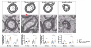
Professor Joutel went on to introduce the relationship between white matter damage caused by ischemic CSVD and abnormal blood-brain barrier function. Using imaging techniques including MRI and DTI, the latest study found that although CADASIL mice showed myelin hypoplasia and axonal structural abnormalities, exogenous tracers could not enter the white matter bundles, suggesting that although the white matter structure was altered, the blood-brain barrier was not significantly impaired.
In the end, Professor Joutel compared the differences between ischemic and hemorrhagic CSVD. Both hemorrhagic CSVD (CoI4a1 mutation) and ischemic CSVD (CADASIL) are genetically heterogeneous and are accompanied by a decrease in arterial smooth muscle cells; hemorrhagic CSVD usually involves hemodynamic changes, whereas ischemic CSVD is associated with decreased smooth muscle cells in the transition zone. Professor Joutel thus surmised that an increase or decrease in parietal cells is the key to differentiating CSVD phenotypes.
Conclusion
Sleep, emotion and cerebrovascular disease are at the very core of human life. When tossing and turning at night, when the mind is in turmoil, when suffering from emotional imbalance, and when cerebrovascular diseases threaten the lives of patients, we feel the invisible constraints and helplessness, and aspire to bring relief to human beings through the power of brain science.
On the long journey to scientific revelations, brain scientists demonstrated both passion and perseverance in their endeavor to overcome scientific hindrances night and day. The rapid advancement of modern technology has opened up new horizons for brain science research. State-of-the-art brain imaging technology, gene sequencing technology, optogenetics and other tools provide unprecedented opportunities for human beings to explore the mysteries of the brain.
In this promising era, we firmly believe that brain science research will bring unprecedented benefits to mankind.

Scan the QR code to watch the replay